
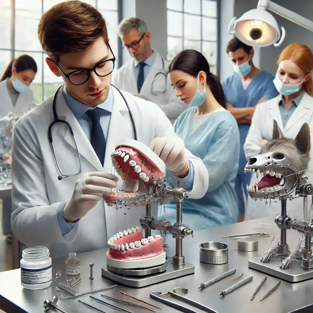
Emergency management for jaw trauma: intensive course with DVM Dr. Ana Nemec, PhD, Dipl. AVDC, Dipl. EVDC, Assistant Prof. on September 13, 2025
Are you looking for a hands-on course to improve your skills in dealing with small animal emergencies and jaw trauma? Then our course on September 13th, 2025 is just right for you! Under the direction of Dr. Ana Nemec , a renowned expert in this field, can expect an exciting and educational day full of lectures and practical exercises.
Course language: English
 Date: September 13, 2025
 Program director/master course leader: DVM Dr. Ana Nemec , PhD, Dipl. AVDC, Dipl. EVDC, Assist. Prof.
 Max. number of participants: 10

 Course location: doc4pets Academy Idar-Oberstein
 ATF hours: expected 7 ATF hours
 Price both days: 999 EUR plus 19% VAT.
Emergency management for jaw trauma: intensive course with Dr. Ana Nemec on September 13, 2025
Are you looking for a hands-on course to improve your skills in dealing with small animal emergencies and jaw trauma? Then our course on September 13th, 2025 is just right for you! Under the direction of Dr. Ana Nemec, a renowned expert in this field, can expect an exciting and educational day full of lectures and practical exercises.
Course content at a glance:
Time: 9:00 a.m. – 6:00 p.m.
Duration: 8 hours
- 9:00 – 9:45 : Oral Emergencies – Gain important insight into the initial care of oral emergencies.
- 9:45 – 10:30 : Maxillofacial Trauma in Cats – Learn all about the unique challenges of jaw injuries in cats.
- 10:30 – 10:45: Coffee break – A short break to recharge your batteries for the practical exercises.
- 10:45 - 11:45 : Practical exercises: Repairing the symphyseal separation in cats - techniques such as cerclage wire and wire-reinforced splints are practiced intensively.
- 11:45 - 12:45 : Practical exercises: Rigid maxillomandibular fixation device (MMF) in cats - Learn how to use and remove these devices.
- 12:45 – 13:30: Lunch break – time to relax and network.
- 1:30 p.m. - 2:30 p.m. : Practical exercises: Functional/semi-rigid MMF in cats and dogs - use of orthodontic brackets and labial buttons.
- 2:30 p.m. – 3:30 p.m .: Practical exercises: Risdon/Stout technique for mandibular fractures – wire-reinforced splints and their removal.
- 3:30 p.m. – 4:30 p.m .: Practical exercises: Repair of the palate separation according to von Langenbeck – Learn an effective technique for restoring the palate.
- 4:30 p.m. – 6:00 p.m .: Practical exercises: Partial pulpectomy and restoration of dog teeth – with interactive evaluation of X-ray images.
Why should you participate?
This course not only provides you with theoretical knowledge, but also numerous practical exercises to refine your skills and develop routine in dealing with complex jaw injuries. Whether you already have experience in this area or would like to refresh your knowledge, this course is ideal for veterinarians who want to specialize in emergency management of jaw trauma.
Secure your place now and benefit from intensive training under the guidance of a leading expert!
Special instructions:
- Prerequisites: Clinical skills in dental radiography, regional anesthesia/analgesia (nerve blocks), periodontal therapy, and tooth extractions are required.
Questions that will be considered in the course include:
What biomechanical principles should be considered when treating jaw fractures in small animals?
Treatment of jaw fractures in small animals requires a deep understanding of the biomechanical principles involved in stabilization and healing. Basically, treatment aims to restore normal occlusion and function of the jaw while ensuring stable fixation. It is crucial to take into account the mechanical forces that act during jaw movements, particularly tension, compression and torsion. In many cases, rigid fixation methods are used, such as intermaxillary fixation (IMF) or the insertion of miniplates, to ensure the anatomical alignment of the fractures. The biomechanical properties of the material used, such as elastic modulus and breaking strength, also play a role in ensuring sufficient stability without affecting bone healing. An additional challenge is maintaining the vascular supply of the fracture segment to avoid delayed healing or osteonecrosis.
What role does preoperative imaging play in the planning of orthodontic procedures in cats and dogs?
Preoperative imaging is an essential part of planning orthodontic procedures in small animals. Detailed radiological findings enable a precise assessment of the type of fracture (e.g. transverse, oblique, comminuted fracture) and the anatomical structures that are affected. While conventional x-rays provide a basic overview, computed tomography (CT) is indispensable in many cases as it allows a three-dimensional reconstruction of the fractures and surrounding tissue. This is particularly important in complex fractures of the upper jaw, lower jaw or temporomandibular joint. In addition, imaging plays a crucial role in identifying associated injuries, such as dentoalveolar fractures or soft tissue complications. A well-designed preoperative plan based on accurate imaging data can optimize surgical techniques, minimize the risk of intraoperative complications, and improve prognosis.
What postoperative complications can occur with orthodontics for pets, and how can they be detected and treated early?
Postoperative complications following orthodontic procedures in cats and dogs can be varied, ranging from mechanical problems to infections. One of the most common complications is malocclusion, which can be caused by inadequate reconstruction of the fracture or incorrect fixation. This often leads to dysfunction of the masticatory system and may require further intervention. Infections, particularly in open fractures or complex soft tissue injuries, may also occur and require immediate antibiotic therapy and thorough wound care. In rare cases, delayed bone healing or non-union may occur, especially if there is insufficient stability or poor vascularization of the affected region. Careful postoperative monitoring, including regular radiological checks, is crucial to detect these complications early. Clinical signs such as persistent pain, swelling, drainage of pus or an unnatural movement pattern of the jaw should be examined immediately so that timely treatment can be initiated.