
- Urinalysis in veterinary medicine: Avoiding errors and optimizing results
- Urinalysis Errors: Inappropriate sample collection methods
- Errors due to inconsistent sample volumes for centrifugation
- Errors due to inappropriate timing of sample collection and analysis
- Errors due to inappropriate use and/or interpretation of reagent strips
- Errors due to failure to take the patient's history into account during interpretation
- FAQs about urinalysis in veterinary medicine
- Summary of urinalysis in veterinary medicine
Urinalysis in veterinary medicine: Avoiding errors and optimizing results
Comprehensive urinalysis is crucial in veterinary medicine to diagnose diseases of the urinary tract as well as various systemic diseases. Although performing urinalysis is inexpensive and can be done in-house with minimal specialized equipment, errors in sample collection, handling, and interpretation are not uncommon. In this article, we highlight the most common sources of error and provide detailed recommendations to ensure accurate and artifact-free results.
(C) https://www.cliniciansbrief.com/article/urinalysis-error-veterinary-medicine-sample-test-results
Urinalysis Errors: Inappropriate sample collection methods
Voided Sample Collection
A released urine sample should ideally be collected directly into a specially designed, tightly sealable container. Second-use containers should be avoided as residues of cleaning agents, food residues or previous contents can falsify the chemical analysis of the urine.
Cystocentesis and sterile catheterization
Cystocentesis (preferred) or sterile catheterization should be used for culture and susceptibility testing of urine samples. Iatrogenic trauma to a blood vessel with the cystocentesis needle can cause a false increase in hemoglobin and erythrocytes in the sediment. Protein can be detected in cases of significant blood contamination.
| Urine collection method | Advantages | Disadvantages | Recommended application |
|---|---|---|---|
| Sample released | Simple, minimally invasive, stress-free for the animal | High risk of contamination, possible falsification of the chemical analysis by impurities | Routine analysis when risk of contamination is low |
| Cystocentesis | Minimal contamination, suitable for culture, precise sampling | Potential trauma from needle, painful for the animal, technical effort | Bacteriological examinations, culture and susceptibility tests |
| Sterile catheterization | Precise sampling, suitable for culture, less invasive than cystocentesis | Risk of urinary tract infections, invasive technology, technical effort | Bacteriological examinations if cystocentesis is not possible |
| Bladder puncture | Avoid contamination from external influences, take samples directly from the bladder | Requires expertise, invasive, risk of bladder injury | When a sterilized sample is required |
| Midstream urine | Reduces risk of contamination compared to exposed samples, easier than catheterization and cystocentesis | More difficult to perform, requires cooperation from the animal, but there is still a risk of contamination | Routine analyzes if the risk of contamination is to be reduced |
| Catheterization in males | Good control over sampling, less painful than females, suitable for routine analysis and emergencies | Potential risk of infection, discomfort to the animal, invasive | In male animals for routine and emergency analyses |
| Catheterization in females | Precise sampling suitable for specific diagnostics and bacterial examinations | Higher risk of infection and injury, invasive and uncomfortable for the animal | For specific diagnostic requirements |
| Urine suction | Allows collection from animals unable or unwilling to urinate, suitable for samples when other methods are not practical | Requires specialized equipment, risk of trauma and stress to the animal | When other collection methods are not feasible |
| Plasmapheresis-assisted collection | Accurate chemical analysis, minimal contamination, suitable for special biochemical studies | Very specialized procedure, requires expertise and specialized equipment, potentially unpleasant for the animal | In specialized biochemical analyses |
| Forced micturition | Suitable for animals with urinary problems or with specific diagnostic requirements, easy to perform | Can be stressful for the animal, high risk of contamination, possible adulteration from external contaminants | For special diagnostic requirements |
Recommendations for choosing the urine collection method
The choice of urine collection method should always be made taking into account the individual patient and specific diagnostic requirements. Each method has its own advantages and disadvantages that must be weighed to achieve the best possible diagnostic accuracy while ensuring the animal's well-being.
Errors due to inconsistent sample volumes for centrifugation
Importance of consistency in centrifugation of urine samples
The consistency of the sample volume is a crucial factor for the accuracy and reliability of urinalysis. A constant volume (ideally 5 mL) should be centrifuged to prepare the sediment. Different volumes result in varying sediment volumes, which can significantly influence the number of cells, crystals and cylinders detected. This can lead to incorrect interpretations and ultimately misdiagnosis.
Effects of different sample volumes
When different volumes of urine are centrifuged, different amounts of sediment are produced. This means that the amount of elements detected in the sample (such as cells, crystals and cylinders) is not directly comparable unless consistent volumes are used. In practice, this means that results can vary greatly depending on how much urine was originally used.
An example to illustrate:
- 5 ml urine : Centrifugation produces approx. 0.5 ml of sediment. This volume allows accurate evaluation and interpretation based on standardized reference values.
- 1 ml urine : Centrifugation produces significantly less sediment (approx. 0.1 ml). The reduced amount of sediment may result in important diagnostic elements being missed or present in insufficient quantities to make an accurate diagnosis.
Effects of different sample volumes on centrifugation
Explanation of the graphic
This graphic illustrates the effects of different sample volumes on the amount of sediment obtained after centrifugation. The left column shows a sample volume of 5 ml urine, which after centrifugation results in a sediment volume of 0.5 ml. The right column shows a sample volume of 1 ml urine, which results in a sediment volume of 0.1 ml. This representation highlights the need for consistent sample volume to achieve reliable and comparable results.
Technique for minimizing artifacts in inconsistent volumes
To minimize artifacts when an adequate volume of urine is not available, all sediment should be resuspended at a constant percentage of the volume of centrifuged urine. For example:
- For 1 ml of centrifuged urine, 0.1 ml of the decanted supernatant should be added to the sediment.
- For 3 ml of centrifuged urine, 0.3 ml of the decanted supernatant should be added.
- For 5 ml of centrifuged urine, 0.5 ml of the supernatant should be added.
This technique ensures that the density of the sediment remains consistent for microscopic examination, regardless of the original sample quantity. This is particularly important to achieve accurate and reproducible results.
Practical recommendations for centrifugation
To achieve the best results, the following recommendations should be followed:
- Use standard volume : Wherever possible, a standard volume of 5 ml urine should be used for centrifugation. This corresponds to the reference values and allows for consistent and accurate interpretation.
- Sediment Reprocessing : If smaller volumes must be used, the sediment should be reprocessed as described above to ensure consistency.
- Calibrated Centrifuge : The centrifuge should be calibrated and maintained regularly to ensure that it operates consistently and reliably.
- Recorded procedures : The exact sample quantity and technique used should be documented to ensure traceability and consistency of results.
Sample volume consistency during centrifugation is critical to the accuracy of urinalysis. By using standardized volumes and techniques to minimize artifacts, the reliability of diagnostic results can be significantly improved. This ultimately contributes to better patient care and more accurate diagnoses.
Errors due to inappropriate timing of sample collection and analysis
Importance of the right time to take samples
The timing of sample collection plays a crucial role in the accuracy and reliability of urinalysis. Various factors such as time of day, previous therapeutic measures and feeding time can significantly influence the composition and properties of urine.
Influence of the time of day
The time of day of sample collection can significantly influence various aspects of urinalysis. For example, a morning urine sample is often more concentrated and better suited to assess renal tubular function. Morning samples provide insight into the kidneys' ability to concentrate urine, which is critical for assessing kidney function. In contrast, later samples may be influenced by the day's food and fluid intake, leading to varying results.
Influence of therapeutic measures
Sample collection should ideally take place before therapeutic measures such as fluid therapy or the administration of diuretics. Such measures can distort the urine specific gravity (USG) and other parameters. For example, fluid therapy may decrease USG by diluting the urine, which may lead to an inaccurate assessment of renal concentrating ability.
Influence of medication and feeding
Medications and dietary intake can also have a significant impact on urinalysis. Antibiotics should not be administered prior to sample collection as they may interfere with urine culture results. Postprandial (after eating) urine samples may show transient alkalinuria (elevated pH), making results difficult to interpret.
Time frame for analysis
For the most accurate results, urine should ideally be analyzed within an hour of collection. Delayed analyzes can lead to numerous artifactual changes:
- Bacterial Growth : Pathogenic or contaminating bacteria can proliferate, resulting in false positive results.
- Changes in chemical composition : Substances such as bilirubin, glucose, and ketones can break down, causing false negative results.
- Changes in physical properties : Urine may have increased cloudiness, odor and pH if not stored correctly.
Storage and handling of samples
If immediate analysis is not possible, urine should be refrigerated in a sealed container away from light. However, storage should not exceed 24 hours. Before analysis, chilled urine should be brought to room temperature to avoid measurement errors. Refrigerated urine may have an increased number of amorphous crystals and show a false increase in urine specific gravity.
Practical recommendations for sample collection and analysis
- Prefer morning samples : Whenever possible, urine samples should be collected early in the morning to accurately assess the kidneys' ability to concentrate.
- Before therapeutic measures : Samples should be collected before any therapeutic intervention such as administration of fluids or medications.
- Rapid analysis : Urine should be analyzed within one hour of collection. If this is not possible, the sample should be refrigerated and analyzed within 24 hours.
- Proper Storage : Refrigerated samples should be brought to room temperature prior to analysis to ensure accurate results.
The timing of sample collection and subsequent analysis is critical to the accuracy and reliability of urinalysis. By taking the above factors into account, the risk of erroneous results can be minimized and diagnostic quality can be significantly improved. Careful planning and execution of sample collection contributes significantly to optimal patient care.
Errors due to inappropriate use and/or interpretation of reagent strips
Importance of reagent strips in urinalysis
Reagent strips, also known as test strips or dipsticks, are an essential tool in urinalysis. They enable rapid and cost-effective semi-quantitative analysis of various urine parameters, including pH, glucose, proteins, ketones, bilirubin and blood. Despite their widespread use in veterinary medicine, there are numerous potential sources of error that can lead to incorrect results and interpretations.
Unreliability of certain parameters
Some parameters on the reagent strips are less reliable for use in animals. For example, tests for leukocyte esterase, nitrites, and urobilinogen developed for human medicine are often unreliable in veterinary medicine. This is due to physiological differences between humans and animals. The presence of leukocytes is better determined by microscopic examination of the sediment. Likewise, a refractometer provides more accurate measurements of urine specific gravity than the equivalent reagent strip fields.
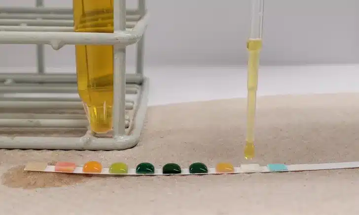
Dip vs. drip method
There are two main methods of using reagent strips: the dip method and the drip method. The choice of method may affect the accuracy of the results.
Dip method
The dip method involves briefly dipping the reagent strip into the urine (approximately one second). This method ensures that all reagent fields are wetted evenly. The disadvantage of this method is that with small sample volumes, complete submersion of the strip can be difficult.
Drip method
The drip method involves applying urine directly to each reagent field using a pipette. This method is particularly useful when only small amounts of urine are available. However, uneven wetting of the reagent fields can lead to inaccurate results.
Dip vs. drip method when using reagent strips
Errors in handling and storing reagent strips
Reagent strips are delicate diagnostic tools whose accuracy can be affected by improper handling and storage. They should always be stored in their original container and protected from moisture, heat and direct light. After opening the container, the lid should immediately be tightly closed again to protect the strips from moisture.
Errors caused by failure to follow the manufacturer's instructions
The manufacturer's instructions should be followed closely to achieve correct results. This includes adhering to the recommended dipping or pipetting times and waiting times until results are read. Too long or too short an exposure time can lead to incorrect results.
Practical recommendations for the use of reagent strips
- Strictly follow manufacturer's instructions : Any deviation from recommended procedures may affect the accuracy of test results.
- Using a Refractometer for USG : Urine specific gravity should preferably be measured with a refractometer as it is more accurate than the corresponding reagent strip fields.
- Regular training of staff : All persons involved in urinalysis should be regularly trained in the correct use and interpretation of reagent strips.
- Use of fresh samples : Urine samples should be as fresh as possible to avoid chemical changes and bacterial growth that could confound test results.
Reagent strips are a valuable tool in urinalysis, but are susceptible to various sources of error. With proper use, storage and interpretation, many of these errors can be avoided. Careful adherence to the manufacturer's instructions and regular training of personnel contribute significantly to the accuracy and reliability of the test results.
Errors due to failure to take the patient's history into account during interpretation
Importance of patient history in urinalysis
Patient history is a crucial factor in accurately interpreting urinalysis results. Without a comprehensive knowledge of dietary habits, medication use and other relevant background information, the results can easily be misunderstood or misinterpreted. A detailed anamnesis helps to distinguish physiological variations from pathological changes and to make a precise diagnosis.
Influence of diet on urinalysis
The animal's diet can have a significant impact on the results of urinalysis. Different foods and diets can change the color, pH, and other chemical properties of urine.
Influence of food
Certain foods such as beets, carrots, blackberries and vitamin C can change the color of urine and lead to pigmenturia. This pigmentation can influence the results of the reagent strip analysis and lead to false-positive or false-negative results.
- Beetroot : May cause reddish urine discoloration, which could be misinterpreted as hematuria.
- Carrots : May cause orange discoloration of urine, which may be confused with bilirubin.
- Vitamin C : High doses can affect the chemical test fields of the reagent strips and cause false-negative results for blood, bilirubin and glucose.
Influence of diet on pH
The pH of urine is strongly influenced by diet. Carnivores such as cats and dogs that are fed a high-protein diet tend to have acidic urine (pH < 7), while herbivores such as rabbits and guinea pigs tend to have alkaline urine (pH > 7).
- High-protein diet : Leads to acidic urine, which must be taken into account when evaluating urinary stones and infections.
- Plant-based diet : Leads to alkaline urine, which can affect the formation of certain crystals and stones.
Influence of medications on urinalysis
Medications, both prescription and over-the-counter, can significantly affect urinalysis results. Accurate knowledge of the animal's medication is therefore essential.
Influence of specific medications
- Diuretics : Increase urine output and may decrease urine specific gravity (USG), making assessment of renal function difficult.
- Antibiotics : May affect urine culture results by killing or inhibiting the growth of pathogenic bacteria.
- Ketamines and other anesthetics : May cause glucosuria and hyposthenuria, which must be taken into account when interpreting results.
- Vitamin C : In high doses it can cause false-negative reactions in the test fields for blood (heme), bilirubin and glucose on the reagent strips.
Practical recommendations to take into account the patient's history
- Comprehensive medical history : Before the urinalysis, a detailed medical history should be taken of the patient, which includes information about diet, medication and previous illnesses.
- Consideration of diet : The animal's diet should always be taken into consideration when interpreting urine results, especially in the case of aberrant pH values and unusual color results.
- Documentation of Medication : All current and recent medications should be documented and considered when interpreting urine results.
- Periodic review of medical history : Medical history should be updated periodically to capture changes in diet or medications that could affect urinalysis.
Failure to take the patient's history into account when interpreting urinalysis can result in significant diagnostic errors. A detailed medical history, including information about diet, medications, and previous medical conditions, is essential for accurate and reliable interpretation of urine results. Careful consideration of these factors can significantly improve diagnostic accuracy and ensure optimal patient care.
FAQs about urinalysis in veterinary medicine
Why is urinalysis important in veterinary medicine?
Urinalysis in veterinary medicine is of great importance because it provides a variety of diagnostic information that helps assess the health status of an animal. It enables the early detection of diseases of the urinary tract such as infections, stones and tumors as well as systemic diseases such as diabetes mellitus, liver and kidney diseases. By analyzing physical (e.g. color, turbidity), chemical (e.g. pH, protein, glucose) and microscopic parameters (e.g. cells, crystals), veterinarians can make a comprehensive health assessment. This allows not only early diagnosis, but also monitoring the progression of the disease and evaluating the effectiveness of therapeutic measures.
What sampling methods are there and which is the best?
There are several methods of sample collection for urinalysis in veterinary medicine , each with its own advantages and disadvantages:
Voided Samples : This method is minimally invasive and easy to perform.
The urine is collected directly into a clean container. The disadvantage is the high risk of contamination from external influences, which can distort the results. Cystocentesis : This involves collecting urine directly from the bladder using a needle.
This method minimizes the risk of contamination and is particularly suitable for bacteriological examinations. However, it is invasive and requires technical skills and sedation or anesthesia of the animal. Sterile catheterization : A catheter is inserted through the urethra into the bladder to collect urine.
This method provides precise sample collection but is invasive and carries the risk of urinary tract infections. Midstream urine : Urine is collected during the midstream, reducing the risk of contamination.
However, this method requires the animal's cooperation and is more difficult to perform. The best method depends on the clinical situation and the diagnostic goal. For general analysis, the blank sample is often sufficient, while for sterile examinations (e.g. culture and susceptibility testing) cystocentesis or sterile catheterization is preferred.What errors can occur when performing and interpreting a urinalysis?
during veterinary urinalysis that affect the accuracy of results:
Inappropriate sample collection : Contaminated or inadequately collected samples can produce false results.
Choosing the wrong sampling method can lead to a bias in the analysis. Inconsistent sample volumes : Different urine volumes during centrifugation result in varying sediment volumes, which affects the interpretation of the cells and crystals.
A standardized volume (ideally 5 ml) should be used. Incorrect timing of sample collection : Samples should preferably be collected in the morning and before therapeutic measures such as fluid therapy or antibiotic administration.
Storage and handling of the sample (e.g. refrigeration) are also crucial. Errors in using reagent strips : Unreliable parameters, improper application (dip vs. drip method), and failure to follow the manufacturer's instructions can lead to erroneous results.
Failure to take into account the patient's history : The animal's diet, medication and previous illnesses must be taken into account during interpretation in order to distinguish physiological variations from pathological changes.How should urine samples be properly stored and handled?
Proper storage and handling of urine samples is critical for veterinary urinalysis to ensure accurate and reliable results.
Here are some important guidelines: Immediate analysis : Ideally, urine should be analyzed within an hour of collection to avoid chemical changes and bacterial growth.
Refrigeration : If immediate analysis is not possible, the sample should be refrigerated in a sealed container.
However, storage should not exceed 24 hours. Bring to room temperature : Chilled urine should be brought to room temperature before analysis to avoid measurement errors.
Refrigerated urine may have an increased number of amorphous crystals and show a false increase in urine specific gravity. Light protection : Urine samples should be protected from direct exposure to light because light can promote the breakdown of certain chemical substances in urine.
Sterile containers : Sterile and clean containers should be used to avoid contamination.Why are regular training and continuing education in the field of urinalysis important?
Regular training and continuing education are of great importance for professionals working
urinalysis in veterinary medicine This ensures that they are familiar with the latest techniques, methods and research. Some of the reasons for the need for continued education include: Updating knowledge : New research and technological developments can improve the methods and techniques of urinalysis.
Regular training helps to integrate these new findings. Quality assurance : Training helps standardize procedures and minimize sources of error, which increases the quality and reliability of analyses.
Patient safety : By applying best practices and current techniques, more accurate diagnoses can be made, thereby improving patient care.
Increased efficiency : Training can also improve the efficiency of analysis processes, leading to faster and more precise results.
Regulatory Requirements : Many regions have regulatory requirements that require regular education and training to maintain licensing and accreditation.
Overall, regular training and further education contribute significantly to optimizing urinalysis in veterinary medicine , which ultimately leads to better diagnosis and treatment of animal patients.
Summary of urinalysis in veterinary medicine
Urinalysis in veterinary medicine is an indispensable diagnostic tool that provides valuable information about the health status of animals. A complete urinalysis in veterinary medicine includes the physical, chemical and microscopic evaluation of urine and helps diagnose urinary tract diseases as well as systemic diseases.
A common error in urinalysis in veterinary medicine is the use of inappropriate sampling methods. Methods such as blank specimens, cystocentesis, and sterile catheterization each have advantages and disadvantages that must be considered when choosing the collection method. Proper technique minimizes the risk of contamination and increases the accuracy of results.
Another error in urinalysis in veterinary medicine arises from inconsistent sample volumes for centrifugation. Different volumes result in varying amounts of sediment, which affects the number of cells and crystals detected. Standardized sediment reprocessing volumes and techniques are critical for consistent and accurate results.
The timing of sample collection is also critical for urinalysis in veterinary medicine . Samples should preferably be collected in the morning and analyzed within one hour. Delays and incorrect storage can lead to artifactual changes that distort the results.
The use and interpretation of reagent strips is another area where errors can occur. In veterinary urinalysis, the manufacturer's instructions should be followed closely to ensure accurate results. The choice between dip and drip methods depends on the sample size, with each method having its own advantages and disadvantages.
Patient history plays a crucial role in urinalysis in veterinary medicine . Diet and medications can significantly influence urine parameters. A comprehensive anamnesis helps to distinguish physiological variations from pathological changes and to make a precise diagnosis.
Overall, urinalysis in veterinary medicine a complex process that requires careful planning and execution. By considering all relevant factors and applying proven techniques, the diagnostic accuracy of urinalysis in veterinary medicine be significantly improved. A thorough history, consistent sample volumes, and proper use of reagent strips are critical to achieving reliable, artifact-free results. Urinalysis in veterinary medicine therefore remains an indispensable tool for optimal patient care.
Urinalysis in veterinary medicine offers numerous diagnostic possibilities, but its accuracy depends heavily on correct implementation and interpretation. It is essential that all professionals involved in urinalysis in veterinary medicine are fully trained and aware of the latest techniques and recommendations.
Proper handling and storage of samples is another critical issue in urinalysis in veterinary medicine . Urine samples should be collected in clean, sterile containers and stored refrigerated if necessary to minimize chemical changes and bacterial growth. Before analysis, chilled urine should be brought to room temperature to ensure accurate measurement results.
A key aspect of urinalysis in veterinary medicine is the microscopic examination of the sediment. This examination allows the identification of cells, crystals, bacteria and other elements relevant to the diagnosis of urinary tract diseases and other health problems. A standardized approach to sample preparation and analysis is crucial to achieve reliable and reproducible results.
The interpretation of urinalysis results in veterinary medicine must always be done in the context of the patient's overall clinical history and current symptoms. Looking at laboratory values in isolation can lead to misinterpretations. Therefore, it is important to correlate urinalysis results in veterinary medicine with other diagnostic information to make an informed diagnosis.
Another challenge with urinalysis in veterinary medicine is that different animal species have different physiological norms. This requires a deep understanding of the species-specific differences and an adjustment of the interpretation criteria accordingly.
Continuous training and research in the field of urinalysis in veterinary medicine contribute to expanding diagnostic options and improving the accuracy of the analyses. New technologies and methods can help to further increase urinalysis in veterinary medicine
In summary, is a versatile and valuable diagnostic tool in veterinary medicine By carefully considering all relevant factors and using proven methods, urinalysis in veterinary medicine help promote animal health and well-being. The constant improvement and adaptation of techniques and procedures are essential to maintain and further develop the high standard of urinalysis in veterinary medicine .
Further information can also be found at: https://www.cliniciansbrief.com/article/urinalysis-error-veterinary-medicine-sample-test-results