
- Abstract: Planning and control of a TPLO surgery
- Introduction Planning and control of a TPLO surgery
- The knee joint – anatomical aspects – Planning and control of a TPLO surgery
- Rupture of the cranial cruciate ligament – incidence, aetiology and pathogenesis – Planning and control of a TPLO surgery
- Diagnosis of anterior cruciate ligament rupture – Planning and control of a TPLO surgery
- Treatment options for anterior cruciate ligament rupture – Planning and control of a TPLO surgery
- Principles of the TPLO – Planning and control of a TPLO surgery
- Preparation of the patient and control of the procedure – Planning and control of a TPLO surgery
- Intra- and postoperative control of TPLO – Planning and control of a TPLO surgery
- Overview of planning and control of a TPLO surgery
- Summary Planning and control of a TPLO surgery
Abstract: Planning and control of a TPLO surgery
The following explanations are largely based on the very good article by Prof. Andrea Meyer-Lindenberg and summarise the key points.
Precise pre-examination and planning prior to surgical procedures, especially tibial plateau leveling osteotomy (TPLO) in veterinary medicine, are crucial to understanding the biomechanical conditions of the knee joint and determining correct surgical procedures. This includes a comprehensive assessment of the joint anatomy, muscle function and body weight of the animal to ensure optimal functionality after surgery. Detailed planning prevents post-operative complications and contributes to the long- term health and mobility of the animal.
Introduction Planning and control of a TPLO surgery
Veterinary medicine has changed considerably in recent decades. In the past, the focus was mainly on the diagnosis and treatment of diseases in farm animals. Since the middle of the 20th century, however, this focus has increasingly shifted towards the care of pets, especially dogs and cats. This change can be seen in parallel to the development in human medicine, which has also increased the demands of pet owners for veterinary care. This has led to the expansion and establishment of advanced diagnostic and therapeutic procedures in veterinary medicine.
The knee joint – anatomical aspects – Planning and control of a TPLO surgery
A dog’s knee joint is analogous to the human knee joint, consisting of the condyles of the femur and the tibia. In contrast to humans, however, the dog’s cranial cruciate ligament plays a more central role in the stability of the knee joint. Injury to this ligament can have serious consequences as it limits the forward movement of the tibia as well as the internal rotation and hyperextension of the knee joint.
A special feature of the dog’s knee joint is the muscle coactivity that supports the stability of the knee joint. Muscles such as the biceps femoris and quadriceps femoris muscles act as agonists and antagonists of the anterior cruciate ligament. The main load during the dog’s stance and weight-bearing phase is borne by the tibial plateau, which has concave and convex shapes depending on the viewing plane. The inclination of the tibial plateau leads to a forward thrust on the cruciate ligaments with each step, especially on the cranial cruciate ligament.
The anatomical and biomechanical alignment of the tibial plateau plays a crucial role in the health and functionality of the knee joint. Measurements of the tibial plateau angle are crucial for the diagnosis and planning of orthopaedic interventions. A correctly measured Tibial Plateau Angle (TPA) is critical to the success of surgical interventions, with the ideal angle for healthy dogs usually being less than 20°.
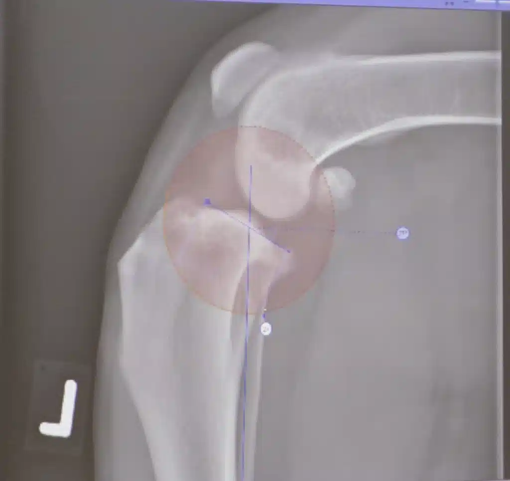
Incorrect radiographic techniques can lead to incorrect measurements which can affect the success of surgery. Measurements to detect deviations of the tibial plateau angle in the lateral plane The angle of inclination of the tibial plateau, also known as the tibial plateau angle (TPA), is determined by special measuring points on a correctly aligned mediolateral X- ray image. The axis of the tibia is shown as a straight line running from the centre of the talus to the highest point of the tibia between the intercondylar tuberosities. The tibial plateau itself is represented by a line that
is orientated to the medial articular surface of the proximal tibia, whereby the boundaries of the tibial plateau are determined by anatomical landmarks.
The Tibial Plateau Slope (TPS) is the angle between the tibial axis and the line of the tibial plateau. To ensure accurate and consistent results, standardised radiographs with correct positioning of the animal are essential. Incorrect radiographic techniques can affect the visualisation of the TPA and lead to measurement errors that can jeopardise the success of surgical procedures. The TPA varies between different breeds of dog and the angulation of the stifle joints can also be different in different breeds. In healthy dogs, the TPA is usually less than 20°.
Measurements to detect axial deviation of the tibia in the frontal plane – Planning and control of a TPLO surgery
Correct diagnosis of axial deviation of the tibia in the frontal plane is crucial for orthopaedic assessment and treatment. There are four main aspects to consider: the height of the apex, the plane of the deviation, the size of the deviation and the direction of the apex. The Centre of Rotation of Angulation (CORA) is a central point defined by the intersection of the proximal and distal axis lines of the femur and tibia.
In order to recognise tibial torsion as well as varus and valgus deformities, special X-ray images are required, which are taken in a caudocranial beam path with a knee joint angle of around 132°. It is important that the patella is positioned centrally in the patellar sulcus and that certain anatomical markers, such as the fabellae and the medial edge of the calcaneus, are clearly recognisable.
Specific points are defined for the measurement to determine the mechanical medial proximal tibial angle (mMPTA) and the mechanical medial distal tibial angle (mMDTA) in the frontal plane. The average mMPTA is approximately 90° and the mMDTA is approximately 93°. No significant differences in these angles were found between the different dog breeds.
(C) https://tierarzt-karlsruhe-durlach.de/tplo-schulung/ – Planning and control of a TPLO surgery
Rupture of the cranial cruciate ligament – incidence, aetiology and pathogenesis – Planning and control of a TPLO surgery
Rupture of the cranial cruciate ligament is a common orthopaedic condition in dogs that usually leads to significant lameness. In contrast to humans, where cruciate ligament ruptures are usually caused by trauma, degenerative changes to the ligament are the more common cause in dogs. These changes can be triggered by a variety of factors, including inflammation, immunological processes, ageing, obesity, instabilities such as patellar luxation and limb malalignment.
A steep slope of the tibial plateau could also predispose to cruciate ligament ruptures. The disease affects both large and small breeds, with larger breeds affected at a younger age and smaller breeds often affected at an older age. However, a clear breed predisposition has not been established.
Partial or complete ruptures of the cranial cruciate ligament lead to increasing instability of the knee joint, which, if left untreated, leads to secondary changes such as osteoarthritis and capsular shrinkage. Cruciate ligament rupture is often associated with damage to the caudal portion of the medial meniscus, whereas primary meniscal lesions are rare in dogs.
Diagnosis is based on medical history, clinical symptoms and palpation findings, whereby the drawer phenomenon and the tibial compression test are important diagnostic tools. X-rays in two planes are essential for diagnosis and treatment planning, and magnetic resonance imaging or arthroscopy can be performed in cases of uncertainty or for a more detailed examination of meniscus lesions.
Numerous surgical procedures have been developed for treatment. These can basically be divided into those that alter the joint statics and those that do not. In medium-sized to large dogs, surgical treatment is often necessary due to the strong forces acting on the joint. The conventional methods, which do not change the joint statics, are not always successful, as the inserted material can tear or lead to renewed instability. For this reason, surgical methods have been developed specifically for medium to large breeds that alter the joint statics in order to neutralise the function of the cranial cruciate ligament and thus enable better stability and healing.
Diagnosis of anterior cruciate ligament rupture – Planning and control of a TPLO surgery
The diagnosis of an anterior cruciate ligament rupture in dogs is based on a careful medical history, clinical symptoms and thorough palpation findings.
Typically, the dog presents from mild to severe lameness, accompanied by a relieving posture and walking on tiptoe. On examination of the stifle joint, it may appear thickened and show instability manifested by the drawer phenomenon, where the tibia can be displaced forwards in relation to the femur.
An additional diagnostic method is the tibial compression test, in which the tibia is compressed between the femur and tarsus during the support phase, typically by the body weight and contraction of the gastrocnemius muscle. This test simulates the forces acting on the knee joint and is used to demonstrate instability by sliding the tibia forwards with the tarsal joint flexed.
X-rays should be taken in two planes to confirm the diagnosis and plan treatment. These can help to identify secondary changes such as increased joint filling or osteoarthritis. In unclear cases or for a more precise assessment of meniscus lesions, magnetic resonance imaging can also be helpful. This allows a more detailed examination of the ruptured cruciate ligament, meniscus and cartilage lesions as well as changes in the bone or surrounding soft tissue. Arthroscopy can also be used for the direct examination of meniscus injuries.
Treatment options for anterior cruciate ligament rupture – Planning and control of a TPLO surgery
There are many surgical methods for the treatment of anterior cruciate ligament rupture in dogs, which can basically be divided into two categories: those that alter the joint statics and those that do not. The latter methods are further subdivided into extracapsular and intracapsular ligament replacement methods. Especially in medium and large dogs weighing more than 20kg, traditional methods that do not alter joint statics are often unsuccessful.
Problems can arise due to tearing of the material used or loosening of the fixation during the healing process, which can lead to renewed lameness and the further development of osteoarthritis or meniscus damage. Due to these challenges, procedures that alter joint statics have been developed specifically for medium to large dog breeds. These approaches consider joint anatomy, muscle function and body weight as an integrated system to replace or neutralise the function of the cranial cruciate ligament, making direct replacement of the cruciate ligament unnecessary.
A prominent example of such a method is the tibial plateau levelling osteotomy (TPLO), in which the caudally inclined tibial plateau is surgically modified in such a way that the anterior displacement of the tibia (cranial tibial translation, CTT) is eliminated. This is achieved by rotating the tibial plateau through a semi-circular saw incision and raising it caudally, thereby changing the biomechanical conditions of the knee joint in such a way that the need for a functioning anterior cruciate ligament is bypassed. After the surgery, the caudal cruciate ligament takes on additional stabilising functions in the knee joint, which means that the joint remains functionally stable even without the anterior cruciate ligament.
Principles of the TPLO – Planning and control of a TPLO surgery
The principles of Tibial Plateau Levelling Osteotomy (TPLO) are based on the alteration of the joint statics and biomechanics of the knee joint. Normally, the axial reaction force during weight bearing of the hind limb is orientated along the longitudinal axis of the tibia. When this force hits the tibial plateau, which slopes from cranial to caudal, it is converted into a compression force (perpendicular to the tibial plateau) and a force directed cranially (parallel to the tibial plateau), which triggers the forward movement of the tibia. In the event of a rupture of the cranial cruciate ligament, this inevitably leads to an undesired cranial displacement of the tibia.
The aim of TPLO is to eliminate this undesirable cranial displacement. This is achieved by raising the caudally inclined tibial plateau using a special corrective osteotomy. This elevation optimises the biomechanical
Inclination of the tibial plateau is changed in such a way that the cranial translation of the tibia (CTT) is cancelled and converted into a gravitational thrust instead. This biomechanical correction leads to a stabilisation of the knee joint, which replaces the original function of the damaged or torn cranial cruciate ligament. Planning on the X- ray image and determination of the required rotation of the tibial plateau
To perform a tibial plateau levelling osteotomy (TPLO), two X-ray images are initially required: one in the mediolateral and one in the craniocaudal beam path. These images make it possible to determine the degree of rotation of the tibial plateau and any necessary axial corrections. Determining these angles is crucial in order to achieve correct repositioning of the knee joint and to change the forces acting on the tibial plateau so that they can be absorbed by the caudal cruciate ligament. The aim is to enable muscular compensation during the loading phase and thereby change the biomechanical conditions in the knee joint in such a way that the anterior displacement of the tibia (cranial tibial translation, CTT) is cancelled.
TPLO aims to rotate and lift the plateau caudally by lifting the caudally sloping tibial plateau using a customised semi-circular saw cut in the proximal tibia. This changes the biomechanics of the knee joint in such a way that functional stability is achieved during the stance phase. Rotating the tibial plateau to around 65° cancels the forward thrust of the tibia, giving the caudal cruciate ligament an additional stabilising role.
It is important that the posterior cruciate ligament is intact, as it is exposed to increased forces after the TPLO surgery. Exceeding the optimal angulation can jeopardise the posterior cruciate ligament. Careful planning and execution of the TPLO is therefore crucial to ensure correct biomechanical adaptation and long-term stability of the knee joint.
Preparation of the patient and control of the procedure – Planning and control of a TPLO surgery
Special instruments and precautions are required to prepare and control the surgical procedure of a tibial plateau levelling osteotomy (TPLO). In addition to the standard instruments and special tools for bone surgery, such as special saw blades and TPLO jigs, specially developed plates are also required to fix the tibial plateau. There are a number of different plate models, with locking plate systems proving particularly effective.
Before the surgery, the patient is surgically prepared and positioned in either the lateral or supine position. After preparation, access is gained to the knee joint, possibly after prior arthroscopy to assess or treat meniscal lesions.
During the surgery itself, a special TPLO jig is used, which is attached to the tibial plateau and the tibial diaphysis using two Kirschner wires. This enables precise control and adjustment of the saw cut during the procedure. The jig is also used to prevent or correct malalignments such as varus or valgus deviations or torsions of the tibia.
After positioning the jig and preparing the tibia, the osteotomy is performed. The exact position and inclination of the saw cut are determined in advance to allow ideal rotation of the tibial plateau. After sawing, the
The tibial plateau is rotated accordingly and fixed in the correct position with a TPLO plate. Care must be taken to ensure that the plate is correctly adapted to the bone and does not damage any joint structures.
Postoperatively, a careful check is carried out using X-rays in the mediolateral and craniocaudal beam path. These images are used to check the saw cut, the rotation of the tibial plateau, the position of the plate and screws and the corrections made.
Intraoperatively, the position of the screws can also be checked fluoroscopically in order to avoid incorrect placement.
Finally, regular radiological checks are important to monitor the healing process and the integration of the osteotomy gap. The TPLO plate normally remains in the body unless complications occur.
Intra- and postoperative control of TPLO – Planning and control of a TPLO surgery
Intraoperative and postoperative monitoring of a tibial plateau levelling osteotomy (TPLO) is crucial to ensure the success of the procedure and minimise potential complications. The correct positioning of the screws can be checked fluoroscopically during the surgery using a C-arm. This check is particularly important as incorrect positioning of the screws – especially when using non-locking plates – is one of the most common problems with TPLO. Screws that protrude into the joint often have to be repositioned or replaced with shorter ones.
When using fixed-angle plates, such as those offered by companies like Synthes, the screw direction is already predetermined, which reduces the risk of incorrect positioning. Nevertheless, a precise check is required, as errors can mainly occur if the plate is positioned incorrectly.
Once the surgery has been completed and the wound has been routinely closed, a further check is carried out using X-rays in mediolateral and craniocaudal views. These images are used to check the position of the plate and screws, the correct position of the saw cut, the correction of the tibial plateau and the axis of the tibia. In addition, the rotation of the tibial plateau, particularly in the caudal region, should be checked again. In the post-operative phase, further radiological checks of the knee joint are recommended to monitor the healing process and the integration of the osteotomy
gap. The plate used normally remains in the body unless specific complications occur.
Overview of planning and control of a TPLO surgery
Overview of Planning and control of a TPLO surgery
Summary Planning and control of a TPLO surgery
- Successful planning and control of TPLO surgery requires a postoperative pain management strategy.
- The planning and control of a TPLO surgery begins with a comprehensive diagnosis and the decision that this treatment is the best option for the patient.
- Thorough planning and control of TPLO surgery includes selecting the appropriate timing for the procedure to ensure optimal recovery.
- Before the surgery, detailed planning, preparation and control of a TPLO surgery is necessary in order to provide all the necessary instruments and materials.
- The planning and control of a TPLO surgery also includes informing the pet owner about the procedure and the subsequent rehabilitation phase.
- During the planning and control of a TPLO surgery, the surgical team is carefully assembled and instructed to ensure maximum efficiency and safety.
- Effective planning and control of a TPLO surgery involves the creation of a detailed surgical plan based on the individual anatomical conditions of the animal.
- The planning and control of a TPLO surgery requires the precise determination of the rotation angle of the tibial plateau in order to achieve the best possible results.
- As part of the planning and control of a TPLO surgery, the anaesthesia is carefully selected and dosed to minimise risks.
- The planning and control of TPLO surgery includes a careful review of patient positioning to optimise access and visibility during the procedure.
- Thorough planning and control of a TPLO surgery ensures that all necessary post-operative measures are prepared to promote a speedy recovery.
- The planning and control of a TPLO surgery includes the provision of resources for immediate post-operative control and care.
- During the planning and control of a TPLO surgery, potential complications are taken into account and plans are developed for their management.
- The planning and control of a TPLO surgery involves close control of blood loss and vital signs during the procedure.
- In the course of planning and control a TPLO surgery, specific aftercare instructions are developed for the pet owner.
- The planning and control of a TPLO surgery includes setting up a follow- up plan to monitor the healing and function of the operated leg.
- Careful planning and control of a TPLO surgery includes the selection of the optimal plate and screws for fixation.
- When planning and control a TPLO surgery, the precise determination of the cutting angle for the osteotomy is crucial.
- The planning and control of a TPLO surgery includes the preoperative determination of the correct rotation of the tibial plateau.
- The need for meniscus treatment is assessed as part of the planning and control of a TPLO surgery.
- The planning and control of a TPLO surgery requires that all persons involved are informed and trained about the specific steps of the procedure.
- The planning and control of a TPLO surgery includes the review and adjustment of physiotherapy measures based on the dog’s recovery.
- Comprehensive planning and control of a TPLO surgery includes regular X-ray checks to confirm the correct position of the implants.
- Planning and control TPLO surgery requires continuous assessment of limb function during the rehabilitation phase.
- During the planning and control of a TPLO surgery, particular attention is paid to the prevention of infections.
- The planning and control of a TPLO surgery involves the careful selection of materials and equipment to ensure the best possible surgical results.
- In the course of planning and control a TPLO surgery, long-term strategies are developed for the health and mobility of the dog.
- Finally, well thought-out planning and control of a TPLO surgery guarantees the highest standards in surgical practice and patient care.
Planning and control of a TPLO surgery will be teached in our courses 2024 – just click here for more information: